
White Matter Lesions in Migraine Patients: What You Need to Know
Cerebral TorqueShare
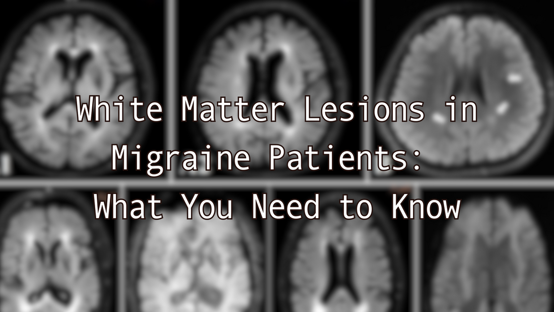
Migraine patients often have MRIs that show white matter lesions (WMLs). It's not uncommon for neurologists to reassure patients that these findings are nothing to worry about, especially in the context of migraine. But what does this really mean? Let's look at the data.
Prevalence
Studies show that 4-59% of people with migraine have WMLs, which is higher than in the general population [1]. This wide range (4-59%) reflects the variability across different studies, which may be due to several factors:
- Differences in study populations (age, gender, migraine type)
- Variations in imaging techniques and MRI protocols
- Different definitions of what constitutes a WML
- The frequency and severity of migraine attacks in the studied population
Types of Brain Changes We Will Discuss
There are two main types of brain changes associated with migraine:
- White matter lesions (WMLs)
- Silent brain infarcts (SBIs)
Potential Causes of WMLs in Patients with Migraine
Several mechanisms might contribute to the development of WMLs in migraine patients:
a) Cortical Spreading Depression (CSD): (I'll link a video here soon when I complete my explanation of CSD)
- CSD is a wave of neuronal and glial depolarization that spreads across the cerebral cortex.
- In migraine, especially migraine with aura, CSD can lead to transient increases in brain tissue blood flow, followed by prolonged periods of reduced blood flow (hypoperfusion).
- Repeated episodes of CSD might lead to small areas of tissue damage over time, potentially manifesting as WMLs [2].
b) Endothelial Dysfunction:
- Migraine patients may show signs of endothelial dysfunction, which affects the inner lining of blood vessels.
- This dysfunction can lead to impaired vascular reactivity and dysregulation of inflammatory pathways.
- Studies have found higher concentrations of markers like asymmetric dimethylarginine (ADMA) and symmetric dimethylarginine (SDMA) in migraine patients with WMLs, suggesting a role of endothelial dysfunction in WML formation [3].
c) Genetic Factors:
- Some genetic predispositions may increase susceptibility to both migraine and vascular brain lesions.
- For instance, mutations in genes like NOTCH3 (associated with CADASIL syndrome) can lead to both migraine and WMLs [4].
Migraine Subtypes and Brain Lesions
- Migraine with aura (MA) seems to have a stronger association with brain lesions compared to migraine without aura (MO).
- Some studies have found a higher prevalence of deep WMLs and brain infarcts in individuals with MA, particularly in older populations [5].
Clinical Significance
The long-term effects of these lesions in migraine patients are still unclear. Most studies don't show a clear connection to cognitive decline or increased stroke risk. However, it's still an area of research and no solid conclusions have been made.
Conflicting Studies
It's important to note that research on the relationship between migraine and brain lesions has produced conflicting findings:
Contradictory Findings:
- While some studies, like Kruit et al. (2004, 2005), found a higher incidence of silent brain infarcts (SBIs) and white matter lesions (WMLs) in migraine patients, others have not replicated these results [6,7].
- The Gaist et al. (2016) study found no significant increase in either SBIs or WMLs in women with migraine [8].
Differences in Migraine Subtypes:
- Some studies suggest that migraine with aura (MA) is more strongly associated with brain lesions than migraine without aura (MO).
- However, not all research supports this distinction. The Monteith et al. (2014) study, for example, found a higher risk of SBIs but not WMLs in migraine patients, regardless of aura status [9].
Age and Gender Factors:
- The Palm-Meinders et al. (2012) longitudinal study showed an increase in WMLs over time in women with migraine, but didn't find a significant increase in SBIs [10].
- Kurth et al. (2011) found more deep WMLs and brain infarcts in older people with MA, suggesting age might play a role in the development of these lesions [5].
Mendelian Randomization Study:
A recent study I posted on r/migrainescience by Huo et al. (2023) used a Mendelian randomization approach to investigate the causal relationship between migraine and white matter lesions [11]. This study found no evidence of a causal relationship in either direction between migraine and WMLs. This suggests that while migraine and WMLs may co-occur, one does not necessarily cause the other.
Key Studies on Migraine and Brain Lesions
| Study | Year | Key Findings |
|---|---|---|
| Kruit et al. | 2004, 2005 | Higher incidence of SBIs in posterior circulation and more deep WMLs in migraine patients, especially women |
| Palm-Meinders et al. | 2012 | Increase in WMLs over 9 years in women with migraine, no significant increase in SBIs |
| Kurth et al. | 2011 | More deep WMLs and brain infarcts in older people with migraine with aura |
| Monteith et al. | 2014 | Higher risk of SBIs in migraine patients, but no increased risk of WMLs |
| Gaist et al. | 2016 | No significant increase in SBIs or WMLs in women with migraine |
| Huo et al. | 2023 | No causal relationship found between migraine and WMLs using Mendelian randomization |
What This Means for You
If your neurologist has told you not to worry about WMLs found on your MRI, their advice is likely based on current medical understanding. Many migraine patients with WMLs don't experience any related symptoms or complications. However, these findings underscore the importance of proper migraine management.
Steps You Can Take:
- Discuss your concerns with your neurologist. They can provide personalized advice based on your specific situation.
- Adhere to your prescribed treatment plan.
- Maintain a healthy lifestyle with regular exercise and a balanced diet.
- Have regular check-ups with your neurologist to monitor any changes over time.
- Learn as much as you can about migraine. The book, Unraveling Migraine, is filled with great, evidence-based information and clinical pearls in regards to migraine treatment.
Remember, while the presence of WMLs might cause alarm, it's important to focus on effective migraine management. As research in this area continues, we'll hopefully gain a clearer understanding of what these brain changes mean for migraine patients.
Until then, your neurologist is your best resource for personalized medical advice.
References:
- Bashir A, Lipton RB, Ashina S, Ashina M. Migraine and structural changes in the brain: a systematic review and meta-analysis. Neurology. 2013; 81:1260-8.
- Yemisci M, Eikermann-Haerter K. Aura and stroke: relationship and what we have learnt from preclinical models. J Headache Pain. 2019; 20:63.
- Erdélyi-Bótor S, et al. Serum L-arginine and dimethylarginine levels in migraine patients with brain white matter lesions. Cephalalgia. 2017; 37:571-80.
- Vahedi K, et al. Migraine with aura and brain magnetic resonance imaging abnormalities in patients with CADASIL. Arch Neurol. 2004; 61:1237-40.
- Kurth T, et al. Headache, migraine, and structural brain lesions and function: population based epidemiology of vascular ageing-MRI study. BMJ. 2011; 342:c7357.
- Kruit MC, et al. Migraine as a risk factor for subclinical brain lesions. JAMA. 2004; 291:427-34.
- Kruit MC, et al. Infarcts in the posterior circulation territory in migraine. The population-based MRI CAMERA study. Brain. 2005; 128:2068-77.
- Gaist D, et al. Migraine with aura and risk of silent brain infarcts and white matter hyperintensities: an MRI study. Brain. 2016; 139:2015-23.
- Monteith T, et al. Migraine, white matter hyperintensities, and subclinical brain infarction in a diverse community: the northern Manhattan study. Stroke. 2014; 45:1830-2.
- Palm-Meinders IH, et al. Structural brain changes in migraine. JAMA. 2012; 308:1889-97.
- Huo, J., et al. (2023). Migraine and white matter lesions: a mendelian randomization study. Scientific Reports, 13(1), 10984.


